A fish veterinarian explains what “Diamond Eye” is
When Jen and I went down our fancy betta rabbit hole in the summer of 2021, we ran across many instances of fishkeepers describing a condition informally known as “diamond eye” in bettas. We also started to see the condition develop in some of the new bettas we’d each acquired, so we wanted to learn more.
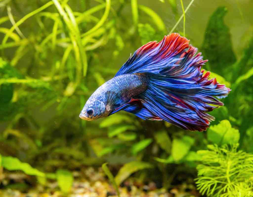
Anecdotally, this condition seems to affect bettas with iridescent coloration near their eyes. As these fish age, the surface of the eye becomes clouded with iridescent color; in advanced cases, the entire surface of the eye can be completely obscured. At the time, I was unable to find any published literature describing and explaining this disease. Fortunately, in the fall of 2022, a group of my colleagues (and my current boss) published a paper about a betta with “diamond eye” that I will break down here (there is a link to their paper at the bottom of this article if you would like to read it yourself).
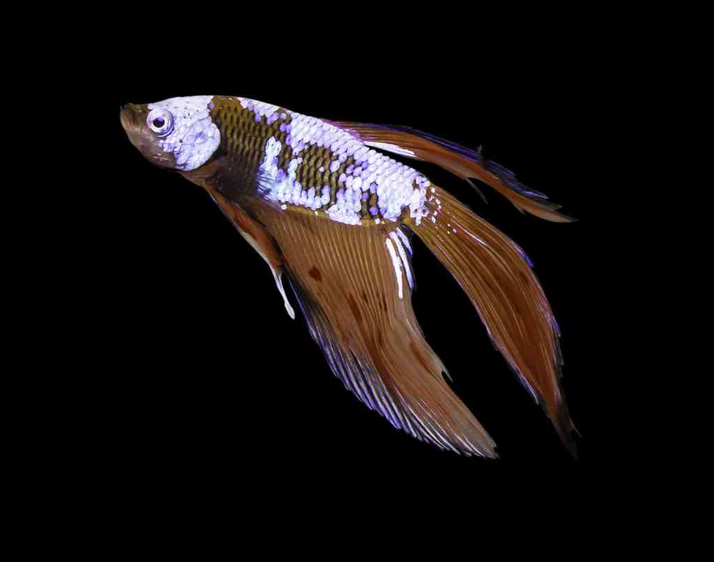
Like many other animals, the outside surface of the fish’s eye is called the cornea. It is composed of epithelial tissue (a type of tissue that makes up the skin and outer surfaces of internal organs) that is normally transparent. This tissue allows light into the eye and protects the inside of the eye from the outside environment. Inside the eye past the cornea is the iris, which is colored (see the picture below of a fish with a white iris) and opens and closes to adjust the amount of light entering the eye.
- Cornea: The transparent front part of the eye that lets in light and protects the eye from the outside.
- Epithelial tissue: This type of tissue is composed of thin sheets of cells; skin and the external surfaces of organs are comprised of epithelial tissue.
- Iris: The muscular ring around the pupil that expands and contracts to let light into the eye; it is the colored part of the eye.
- Chromatophore: Cell with pigment that is located in the skin and gives fish their color.
- Iridophore: Pigment cell in fish’s skin that contributes to metallic and iridescent colors.
- Leucophore: Pigment cell in fish’s skin that contributes to white color.
- Melanophore: Pigment cell in fish’s skin that contributes to black color.
- Chromatophoroma: A tumor of pigment cells (chromatophores).
- Uveitis: Inflammation inside the eye.
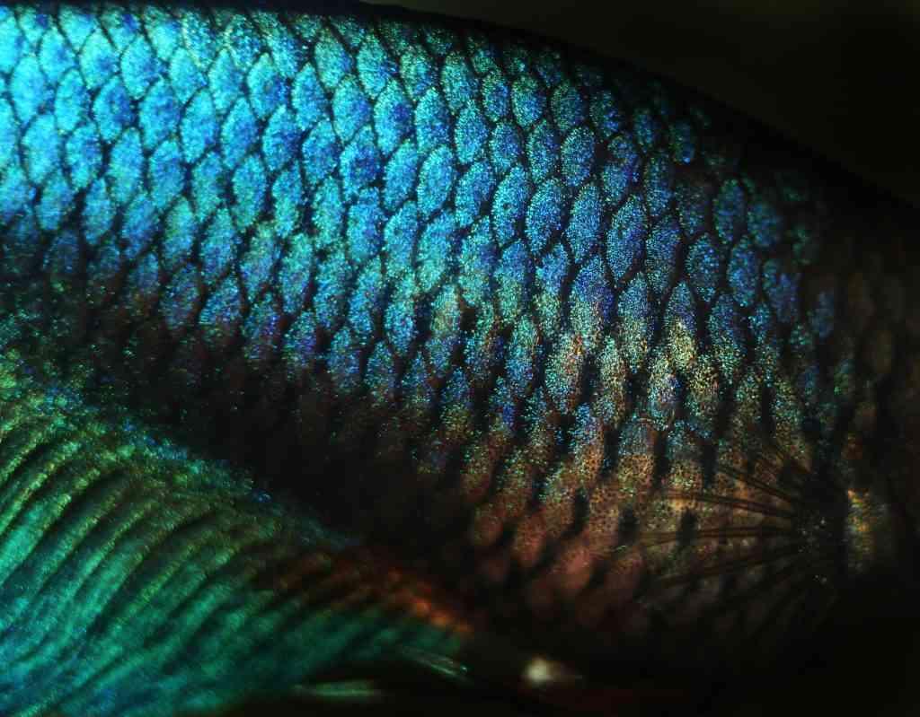
The microanatomy of fish skin is also important for understanding this scientific paper. Fish have a thin layer of epithelial tissue that covers their scales. Chromatophores, the cells that cause a fish’s color, live in this epithelial tissue. There are a number of different chromatophores, and each is responsible for a different color. Metallic and iridescent colors are created by iridophores. White colors are created by leucophores. Black coloration is created by melanophores.
The betta featured in this paper was brought to a fish veterinarian because it had both “diamond eye” and a mass growing on its tail. The fish veterinarian anesthetized the fish and took a small sample from the tail mass and submitted it to a diagnostic laboratory, where it was diagnosed as a tumor of chromatophores. These types of tumors are common in betta fish. The tumor could not be completely removed and was negatively affecting the betta’s quality of life, so the betta was euthanized ten weeks after its initial vet visit. After euthanasia, the entire body was submitted to a diagnostic laboratory for evaluation.
A veterinary pathologist evaluated the affected eye histopathologically and observed inflammation of the cornea and the presence of chromatophores, which are not present in the cornea of a normal fish’s eye. This betta’s eye also had anterior uveitis (inflammation in the chamber of the eye between the cornea and the iris).
The prognosis for fish that develop this condition is poor, and little can be done to help the affected fish. And this is not just a superficial or cosmetic condition. Anterior uveitis can be very painful in other species, and fish with anterior uveitis probably have pain and decreased quality of life. Because bettas are very small, treatments and interventions like surgery are hard to perform effectively and without significant risk and harm to the fish.
One question not answered by this paper is why betta fish develop “diamond eye,” but there is a suspicion among fish veterinarians that this is a consequence of breeding for certain colors and patterns. Future research on the cause and predisposing factors for this condition would be helpful for fishkeepers who wish to responsibly breed and purchase bettas.